TNFRSF10A
TNFRSF10A是一種與腫瘤壞死因子超家族相關的蛋白,其全名為腫瘤壞死因子受體超家族成員10A。該蛋白也被稱為TRAIL-R1、CD261、DR4和APO2。TNFRSF10A在細胞凋亡過程中扮演重要角色,能夠與TRAIL(腫瘤壞死因子相關凋亡誘導配體)結合,觸發細胞凋亡信號通路。它在多種疾病中發揮作用,如癌癥和自身免疫性疾病。目前,針對TNFRSF10A的藥物研發正在進展中,包括開發能夠特異性結合并抑制該蛋白的抗體藥物,以用于治療相關疾病。
熱銷產品
驗證數據
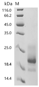
(Tris-Glycine gel) Discontinuous SDS-PAGE (reduced) with 5% enrichment gel and 15% separation gel.
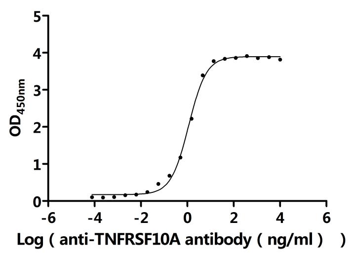
Activity
Measured by its binding ability in a functional ELISA.Immobilized Human TNFRSF10A at 2 μg/ml can bind anti-TNFRSF10A recombinant antibody(CSB-RA023964MA1HU).The EC50 is 1.038-1.260 ng/mL.
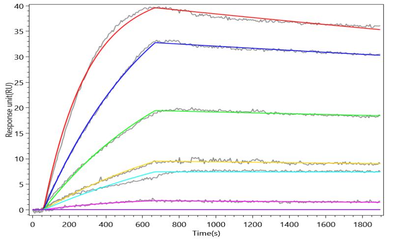
Activity
TNFRSF10A Recombinant Monoclonal Antibody(CSB-RA023964MA1HU) captured on Protein A Chip can bind Human TNFRSF10A with an affinity constant of 0.851 nM as detected by MetaSPR Assay (WeSPRTM 200).
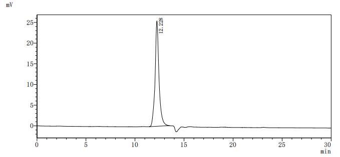
The purity of TNFRSF10A was greater than 95% as determined by SEC-HPLC
TNFRSF10A Antibody (CSB-PA023964LA01HU)
驗證數據
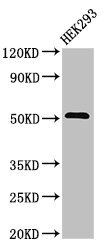
Western Blot
Positive WB detected in: HEK293 whole cell lysate
All lanes: TNFRSF10A antibody at 3.2μg/ml
Secondary
Goat polyclonal to rabbit IgG at 1/50000 dilution
Predicted band size: 51 kDa
Observed band size: 51 kDa
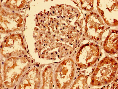
IHC image of CSB-PA023964LA01HU diluted at 1:400 and staining in paraffin-embedded human kidney tissue performed on a Leica BondTM system. After dewaxing and hydration, antigen retrieval was mediated by high pressure in a citrate buffer (pH 6.0). Section was blocked with 10% normal goat serum 30min at RT. Then primary antibody (1% BSA) was incubated at 4°C overnight. The primary is detected by a biotinylated secondary antibody and visualized using an HRP conjugated SP system.
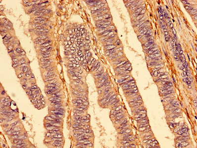
IHC image of CSB-PA023964LA01HU diluted at 1:400 and staining in paraffin-embedded human colon cancer performed on a Leica BondTM system. After dewaxing and hydration, antigen retrieval was mediated by high pressure in a citrate buffer (pH 6.0). Section was blocked with 10% normal goat serum 30min at RT. Then primary antibody (1% BSA) was incubated at 4°C overnight. The primary is detected by a biotinylated secondary antibody and visualized using an HRP conjugated SP system.
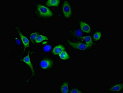
Immunofluorescent analysis of A549 cells using CSB-PA023964LA01HU at dilution of 1:100 and Alexa Fluor 488-congugated AffiniPure Goat Anti-Rabbit IgG(H+L)
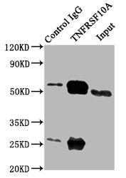
Immunoprecipitating TNFRSF10A in 293 whole cell lysate
Lane 1: Rabbit control IgG (1μg) instead of CSB-PA023964LA01HU in 293 whole cell lysate.
For western blotting, a HRP-conjugated Protein G antibody was used as the secondary antibody (1/2000)
Lane 2: CSB-PA023964LA01HU (6μg) + 293 whole cell lysate (500μg)
Lane 3: 293 whole cell lysate (10μg)
TNFRSF10A Antibodies
TNFRSF10A for Homo sapiens (Human)
| 產品貨號 | 產品名稱 | 種屬反應性 | 應用類型 |
|---|---|---|---|
| CSB-PA199651 | TNFRSF10A Antibody | Human | ELISA,WB |
| CSB-PA002191 | TNFRSF10A Antibody | Human,Monkey | WB, IF, ELISA |
| CSB-PA023964LA01HU | TNFRSF10A Antibody | Human | ELISA, WB, IHC, IF, IP |
| CSB-PA023964LB01HU | TNFRSF10A Antibody, HRP conjugated | Human | ELISA |
| CSB-PA023964LC01HU | TNFRSF10A Antibody, FITC conjugated | Human | |
| CSB-PA023964LD01HU | TNFRSF10A Antibody, Biotin conjugated | Human | ELISA |
TNFRSF10A Proteins
TNFRSF10A Proteins for Homo sapiens (Human)
| 產品貨號 | 產品名稱 | 來源 |
|---|---|---|
| CSB-YP023964HU1 CSB-EP023964HU1 CSB-BP023964HU1 CSB-EP023964HU1-B |
Recombinant Human Tumor necrosis factor receptor superfamily member 10A (TNFRSF10A), partial | Yeast E.coli Baculovirus In Vivo Biotinylation in E.coli |
| CSB-CF023964HU | Recombinant Human Tumor necrosis factor receptor superfamily member 10A (TNFRSF10A) | in vitro E.coli expression system |